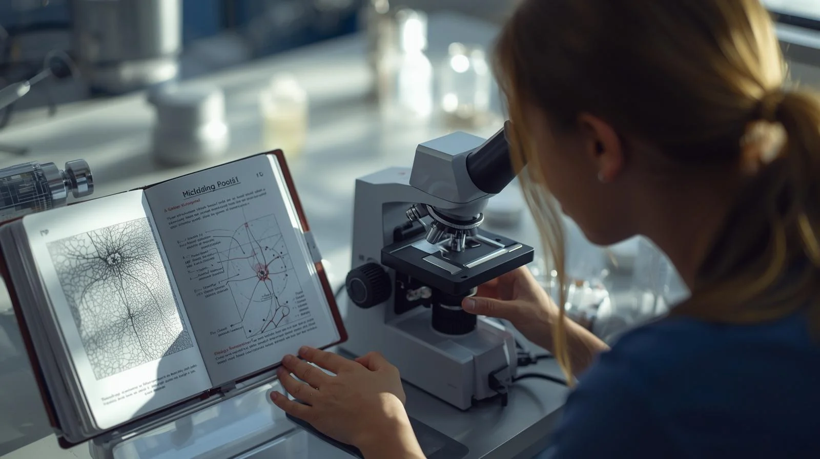
The study of mitosis in onion root tip is a common biology practical that helps students observe cell division stages under a microscope. It demonstrates how a single cell divides into two identical cells through mitosis.
Did you know a single plant cell can make a copy of its genetic blueprint and split into two in under an hour? This extraordinary process is key to all growth in living things. I’m here to guide you through the study of mitosis in onion root tip tissue. It’s a classic experiment that shows how cells divide.

My 2026 guide offers the latest, detailed steps for your lab work. By following these steps, you’ll deeply understand how cells copy their genetic material. You’ll also learn to make slides that show the cell division stages clearly under a microscope.

Key Takeaways
- Understand the fundamental stages of cellular replication.
- Learn the precise steps for preparing plant tissue slides.
- Identify distinct phases like prophase, metaphase, anaphase, and telophase.
- Master the use of laboratory stains to enhance visibility.
- Gain practical skills for high-quality biological observation.
Understanding the Biological Significance of Mitosis
Learning about mitosis is key to understanding plant biology. It’s the fundamental mechanism that helps plants grow by adding cells. By studying mitosis in onion root tips, we see how plants grow and fix themselves at a tiny scale.
The Role of Cell Division in Plant Growth
Plant growth doesn’t happen all over. It focuses on special areas called meristems. These spots have actively dividing cells that help make new leaves, stems, and roots.
Mitosis makes sure each new cell gets the same genetic info. This is key for keeping the plant’s structure strong. Without it, plants wouldn’t grow or work right.
Why Onion Root Tips are Ideal for Cytological Research
I picked onion root tips for their role in cell studies. They have lots of cells that divide fast and are easy to study. This makes them perfect for seeing onion root tip chromosomal behavior.
The root tip’s shape is great for making slides. It lets us see a monolayer view under the microscope. This makes studying mitosis in onion root tips a top choice for anyone interested in plant cell behavior.
Essential Laboratory Equipment and Reagents
Getting clear results in my experiment depends on the quality of my equipment and reagents. I make sure my workspace is organized. I also make sure every tool is ready before I start observing onion root tip mitosis.
Microscope Specifications for High-Resolution Imaging
I use a compound light microscope to see cell division details. It needs at least 400x magnification to clearly show onion root tip chromosomal behavior. This level of magnification is essential for capturing the fine details of cell division.
Chemical Requirements for Fixation and Staining
Specific chemicals are needed to prepare the tissue and make structures visible. I use standard reagents to make chromosomes stand out. Here’s what I need for my lab kit:
- Acetocarmine stain: This is vital for highlighting the chromatin and chromosomes.
- Hydrochloric acid (1M): Used for the hydrolysis of the cell wall to soften the tissue.
- Fixative solution: Typically a mixture of ethanol and acetic acid to preserve cellular structure.
- Distilled water: Required for rinsing the root tips between chemical treatments.
Handling Acetocarmine and Hydrochloric Acid Safely
I handle these chemicals with great care because they can be dangerous. I always wear a lab coat, gloves, and safety goggles. Safety is my top priority when working with acidic solutions.
After I finish, I make sure to dispose of chemical waste properly. I never pour concentrated acids down the drain without neutralizing them first. By following these steps, I keep my research environment safe for studying onion root tip chromosomal behavior.
Preparing the Onion Root Tips for Observation
Getting clear microscopic images starts with how you grow your onion roots. The quality of your slides depends on the overall health of the tissue. Focus on the meristematic zone for samples full of active cells.
Cultivating Healthy Roots in Laboratory Conditions
Start by placing a fresh onion bulb over a beaker of clean water. Make sure only the basal plate touches the water to avoid rot. In three to four days, you’ll see vigorous root growth from the base.
This setup helps roots grow long enough for easy handling. Keep your lab at a steady temperature for consistent growth. If the water gets cloudy, change it to keep things clean and healthy for onion root tip mitosis.
“Nature does not hurry, yet everything is accomplished. In the laboratory, patience with your specimens is the first step toward scientific discovery.”
Timing the Harvest for Maximum Mitotic Activity
Timing is key to catch the onion root tip mitotic phase in action. Cell division peaks in the early morning. Harvesting then gives you a lot of cells in different division stages.
Harvesting too late means fewer cells dividing. Use a sharp blade to cut off the terminal 5mm of the root tip. This part has the most meristematic cells for your study.
| Growth Factor | Optimal Condition | Impact on Results |
|---|---|---|
| Water Quality | Distilled or Dechlorinated | Prevents tissue decay |
| Light Exposure | Indirect/Dim Light | Promotes root elongation |
| Harvest Time | Early Morning | Increases mitotic index |
By following these steps, you set up your research for success. Proper preparation means every slide will show the details of cellular reproduction clearly. Have a look of Kota Notes for NEET: Free PDF Download (Physics, Chemistry, Biology) 2026 Guide to learn more for your upcoming exams.
The Step-by-Step Procedure for Slide Preparation
To see the onion root tip mitotic phase clearly, I use specific lab steps. These steps help keep the cells’ details intact for viewing under a microscope.

Fixing the Root Tip Tissue
I start by soaking the root tips in Carnoy’s fluid. This step is crucial because it stops the cells from living and keeps their structures intact. It makes sure the chromosomes stay in place for later study.
Hydrolysis and Softening of the Cell Wall
Plant cells have tough walls that hide their details. To fix this, I use hydrolysis by heating the tips in hydrochloric acid. This softens the walls, making it easy to flatten the tissue later.
Staining Techniques for Chromosomal Visibility
After softening, I stain the tissue with acetocarmine or aceto-orcein. These stains make the chromosomes stand out against the rest of the cell. This contrast is essential for seeing the cell division in onion root tip stages.
Squashing the Tissue for a Monolayer View
Then, I put the stained root tip on a slide and press it with a coverslip. This squashing spreads the cells into a single layer. A single layer is key for seeing the chromosomes clearly during the onion root tip mitotic phase.
| Reagent | Primary Function | Duration |
|---|---|---|
| Carnoy’s Fixative | Preserves cell structure | 24 Hours |
| Hydrochloric Acid | Softens cell walls | 5-10 Minutes |
| Acetocarmine Stain | Highlights chromosomes | 2-3 Minutes |
Detailed Study of Mitosis in Onion Root Tip Phases
Looking through the eyepiece, you see life’s basic mechanics in action. Mastering the stages of cell division in onion root tip studies is very rewarding. By following a systematic approach, you can confidently categorize every cell you observe.
“The cell is the basic unit of life, and its division is the heartbeat of biological growth and repair.”
Identifying Prophase Characteristics
In prophase, the first stage of mitosis, chromatin starts to condense into visible threads. Look for the disappearance of the nucleolus and the breakdown of the nuclear envelope. These signs show the cell is getting ready to divide its genetic material.
Observing Metaphase Alignment
Metaphase is the most striking phase in onion root tip cell division research. You’ll see chromosomes align perfectly in the middle of the cell. This alignment is crucial for ensuring each daughter cell gets the same chromosomes.
Tracking Anaphase Chromosomal Separation
In anaphase, sister chromatids pull apart. They move to opposite poles of the cell, guided by spindle fibers. This rapid separation shows the mitotic apparatus’s mechanical efficiency.
Recognizing Telophase and Cytokinesis
In telophase, chromosomes reach the poles and start to decondense. You’ll see a new nuclear membrane form around each set of chromosomes. Then, cytokinesis happens as a new cell wall forms, dividing the cytoplasm into two daughter cells.
Analyzing Chromosomal Behavior Under the Microscope
Doing a onion root tip microscope study means spotting different division stages. It’s exciting to see how genetic material gets organized. Adjusting focus and light helps show the cell nucleus’s hidden details.
Interpreting Chromatin Condensation Patterns
At the start of cell division, chromatin is a loose network. As division gets closer, it coils into tight structures. The nucleus gets darker and clearer as it condenses.
Careful observation is key to tell early prophase from later stages. In the early stages, chromatin looks like a messy ball. As it tightens, chromosomes show up clearly under the microscope.
Distinguishing Between Sister Chromatids and Daughter Chromosomes
In onion root tip cell division research, spotting separation is vital. Before anaphase, you see sister chromatids joined at the centromere. These look like two arms connected at a central point.
When the centromere splits, they become daughter chromosomes. These move quickly to opposite cell poles. Watching this helps understand how genes are spread.
| Feature | Sister Chromatids | Daughter Chromosomes |
|---|---|---|
| Location | Joined at centromere | Moving to opposite poles |
| Timing | Prophase to Metaphase | Anaphase to Telophase |
| Appearance | X-shaped structure | V-shaped or rod-like |
| Genetic Status | Identical copies | Separated genetic units |
Calculating the Mitotic Index in Onion Root Tip
Calculating the mitotic index lets me turn my observations into real data. This quantitative analysis helps me measure how fast cells divide in the root tip. It’s key to my onion root tip microscope study, showing how active the cells are.
Mathematical Formula for Mitotic Index
To find the mitotic index, I use a simple formula. I count all cells in a field and see how many are dividing.
The formula is:
Mitotic Index = (Number of cells in mitosis / Total number of cells observed) × 100
This makes it easy to compare different samples. It’s essential to count enough cells for accurate results in my onion root tip microscope study.
Significance of the Index in Cell Cycle Research
The mitotic index is key to understanding plant growth. A higher index means cells are dividing quickly in the meristematic area.
This info helps me understand onion root health and growth. Learning this calculation makes my lab reports more detailed and accurate.
Common Challenges and Troubleshooting Tips
Even experts sometimes struggle with sample preparation for microscopic analysis. When calculating the mitotic index in onion root tip, technical problems can pop up. But, most of these issues can be fixed with a few tweaks to your lab technique.

Addressing Poor Staining Results
If your chromosomes look faint or lack contrast, it’s probably the stain’s fault. Check the stain’s concentration, like acetocarmine or orcein, to make sure it’s good. Increasing the immersion time a bit can help the dye get into the cells better.
If the stain is too dark, it might hide the details you need. Try rinsing with a weak acid solution to remove extra dye. Keeping your timing consistent is key to making high-quality slides.
Correcting Over-Squashing and Tissue Damage
Over-squashing can damage cells and distort their shape. Too much pressure can break cell walls, causing cytoplasm to leak and chromosomes to scatter. Use firm, vertical pressure without moving the slide to keep cells intact.
If your tissue gets damaged, use a fresh root tip and apply less pressure during squashing. Keeping a clean, even layer is vital for an accurate mitotic index in onion root tip count. Proper technique helps keep each phase distinct and visible. For best parental care for your children, explore How Can Parents Encourage Their Children to Develop Positive Learning Habits? 2026.
Managing Microscope Focus Issues
Blurry images often come from uneven slides or wrong cover slip placement. Make sure the cover slip is pressed flat for a uniform focus. If focus is a problem, check that your lens is clean.
Adjust the focus slowly to get a clear view of the cells. This precision is critical for identifying phases in mitotic index in onion root tip research. Here’s a quick guide to common problems and how to solve them.
| Problem | Likely Cause | Recommended Solution |
|---|---|---|
| Faint Staining | Insufficient dye time | Increase staining duration |
| Tissue Damage | Excessive pressure | Apply gentle, vertical force |
| Blurry Focus | Uneven cover slip | Ensure flat, clean mounting |
| Low Contrast | Old stain solution | Prepare fresh reagent |
Safety Protocols and Laboratory Best Practices
Keeping your lab safe is key when studying the mitotic index in onion root tip. Make sure your workspace is clean and tidy before starting. This keeps you safe and ensures your results are accurate.
Personal Protective Equipment Requirements
Wearing the right Personal Protective Equipment (PPE) is a must in the lab. A lab coat protects your skin and clothes from spills. Safety goggles shield your eyes from splashes when working with chemicals.
Gloves are also vital for your safety. I use nitrile gloves to block chemicals. Always change your gloves if they get damaged or dirty.
| Equipment | Primary Function | Safety Benefit |
|---|---|---|
| Safety Goggles | Eye Protection | Prevents chemical splashes |
| Nitrile Gloves | Hand Protection | Avoids skin absorption |
| Lab Coat | Body Protection | Shields clothing from stains |
Proper Disposal of Chemical Waste
Disposing of chemical waste correctly is essential. Never dump chemicals down the sink. It’s harmful to the environment and breaks safety rules. Instead, use designated containers for hazardous waste.
After calculating the mitotic index in onion root tip, sort your waste properly. If unsure, check the safety data sheet (SDS) or ask your supervisor. Following these steps keeps your research safe and compliant.
Integrating Matrix Sikar Notes for Advanced Learning
To do well in biology exams, it’s key to link what you see under the microscope with study materials. Mixing lab work with academic resources boosts your memory of complex topics. Using matrix sikar notes helps you understand the onion root tip cell cycle better.
Key Concepts for Competitive Biology Exams
Exams test your skill in linking microscopic images to biochemical stages. The matrix sikar notes break down these stages, helping you catch all the details. Learning these concepts makes you quicker and more accurate in identifying stages like prophase or anaphase.
These notes focus on the proteins and checkpoints that control division. Studying them deepens your understanding of the precision needed in cell replication. This knowledge is crucial for high scores on exams.
Applying Theoretical Knowledge to Practical Observations
Linking your lab work to study materials is key to mastering the subject. When looking at a slide, try to match what you see with your matrix sikar notes. This method strengthens your memory of the onion root tip cell cycle.
If you see a cell in metaphase, relate the chromosome alignment to your study guide. This turns a simple observation into a deep learning experience. Regularly linking theory to practice builds your confidence in biology exams.
Conclusion
Looking through a microscope changes how we see life. It turns complex ideas into something real. By studying the onion root tip cell cycle, we learn how living things grow and fix themselves.
I suggest you try these methods in your own lab work. It will help you get better at analyzing things. Making slides carefully and watching closely are key to seeing cell division clearly.
This hands-on learning is essential for studying genetics and botany. It helps you feel sure when working with tiny biological samples. You’ll also understand complex cell information better.
Keep working on your staining and squashing techniques for better results. Your hard work will help you understand the basic life processes. I’m excited to see how you use these skills in your future studies.
FAQ – study of mitosis in onion root tip
Why do I use onion root tips for studying mitosis?
Onion root tips are perfect for studying mitosis because they have lots of cells that divide quickly. This makes them great for seeing all stages of cell division under a microscope. It’s easier to observe mitosis in these cells than in other plant tissues.
How do I calculate the mitotic index in onion root tip preparations?
To find the mitotic index, I count cells in mitosis and divide by the total cells in the area. This method helps me measure how fast cells are growing and dividing. It’s key for studying onion root tip cell division.
What is the function of acetocarmine and hydrochloric acid in the onion root tip mitosis procedure?
Hydrochloric acid softens the tissue by breaking down pectin in cell walls. Then, I use acetocarmine to stain DNA. This stain helps see chromosomes clearly under a microscope, making it easier to study chromosomal behavior.
When is the best time to harvest roots for studying mitosis in onion root tips?
Mitosis activity peaks in the mid-morning, around 8:00 AM to 10:00 AM. Harvesting roots during this time ensures more cells are in active phases like metaphase and anaphase. This is important for detailed cell division research.
How can Matrix Sikar notes help me understand the theoretical aspects of this experiment?
Matrix Sikar notes are very helpful. They connect abstract concepts to real observations. These notes help you understand chromosomal behavior and link your microscope findings to biological theories needed for exams.
What should I do if my onion root tip mitotic phase observation is obscured by overlapping cells?
Overlapping cells mean the squashing wasn’t enough. Apply firm, vertical pressure to the coverslip to make a thin layer. Refer to Matrix Sikar notes for tips on softening the tissue during hydrolysis.
Are there specific safety protocols I must follow during an onion root tip microscope study?
Safety is very important. Always wear gloves and safety goggles when handling chemicals like acetocarmine and hydrochloric acid. Also, dispose of chemical waste properly to keep the lab safe and clean.